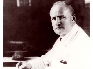
Berger Hans biography
Date of birth : 1873-05-21
Date of death : -
Birthplace : Thuringia, Germany
Nationality : German
Category : Science and Technology
Last modified : 2010-05-26
Credited as : Psychiatrist, Electroencephalogram (EEG), Professor Emeritus in Psychology
0 votes so far
Hans Berger was born in May 21st, 1873, in Neuses near Coburg, Thuringia, Germany. He was the son of the physician Paul Friedrich Berger and Anna Ruckert. One of his grandfathers was Friedrich Ruckert (1788-1866), professor of Oriental languages and a much-quoted poet, whose work is still alive.
Berger graduated from the Gymnasium in Coburg and then entered the University of Jena. After one semester in astronomy he transferred to medicine. In 1897 he received his doctorate and became assistant to Otto Ludwig Binswanger (1852-1929) at the university’s psychiatric clinic. Oscar Vogt and Korbinian Brodmann (1868-1918) were also assistants in the same clinic and encouraged young Berger to join in their work on cerebral localization.
Hans Berger was habilitated in Jena in 1901 and was appointed ausserordentlicher professor in 1906, 1912 physician-in-chief at the clinic, and in 1919 became director of the clinic and succeeded Binswanger in the chair of psychiatry and neurology. He was rector of the university 1927-1928, and prorector 1935 to 1938, when he was emerited. His appointment at Jena was the beginning of an illustrious scientific career. Hans Berger lived in Jena for 41 years until he became Professor Emeritus in Psychology in 1938.
The central theme in Berger’s work was the search for the correlation between objective activity of the brain and subjective psychic phenomena. In his work on blood circulation in the brain (1901) he described his efforts to gain insight into this correlation through plethysmographic registration of the brain pulsations. He investigated the influence of the heartbeat, respiration, vasomotor functions, and position of the head and body on brain pulsations, which were measured through an opening, made by trephination, in the skull.
After series of disappointing experiments measuring the blood circulation and temperature of the brain during the first two decades of the century, Berger following his return from World War I devoted himself mainly to the measurement of the brain’s electrical activity. In 1902 he had taken measurements of electrical activity above skull defects with the Lippmann capillary electrometer (Gabriel Lippman, 1845-1921), and later with the Edelmann galvanometer.
In 1910, however, Berger mentioned in his journal that the results of these measurements were not satisfactory. Therefore, until 1925 he followed two methods of research: stimulation of the motor cortex through a defect in the skull, measuring the time between stimulus and contralateral motor reaction, and registration of the spontaneous potential difference of the brain surface.
After 1925 Berger no longer used the stimulation method. He specialized, with ever increasing skill, in registering the spontaneous fluctuations in electrical potential that could be recorded through the skull from the cortex. In his first publication on electroencephalography (1929), he called July 6, 1924 the date of discovery of the human electroencephalogram, which he called “Elektroenkephalogramm.”
He did this not only in normal subjects but also in the brain-injured, thereby laying the foundation for the application of the technique to clinical technology. In the following year, using a Siemens double-coil galvanometer, he found a decrease in activity on sensory stimulation – thus duplicating the results obtained by Beck and Pravdich-Neminski in animals and he also found the counterpart of two of Pravdich-Neminski’s categories of waves, the alpha and beta ranges.
Electroencephalography (EEG) uses electrodes (made of lead, zinc, platinum, etc.) attached to the intact skull and connected to an oscillograph. The result is a visual picture of brain wave rhythms. Berger made seventy-three EEG recordings from his fifteen-yeasr-old son, Klaus.
The first frequency he encountered was the 10-hertz range, (8 to 12 Hz) which at first was called the Berger rhythm, currently called Alpha rhythm brain wave. After five years of investigation and re-examination of his results, he published his findings. He reported that the brain generates electrical impulses or ‘brain waves’. The brain waves changed dramatically if the subject simply shifts from sitting quietly with eyes closed (short or alpha waves) to sitting quietly with eyes opened (long or beta waves).
Furthermore, brain waves also changed when the subject sat quietly with eyes closed, “focusing” on solving a math problem (beta waves). That is, the electrical brain wave pattern shifts with attention. The publication of Hans Berger’s “On the Electroencephalogram of Man” in 1929 changed neurophysiology forever. Hans Berger thus earned the recognition of “Father of Electroencephalography.”
In developing electroencephalography, Berger was fully aware that Richard Caton (1842-1926), a Liverpool surgeon, had succeeded in 1875 in measuring electrical potentials on the exposed cortex of experimental animals (rabbit and monkey), and that he was thus the discoverer of the electrical activity of the brain. Berger also knew about the further successes along this line achieved by the Polish physiologist Adolf Beck (1863-1939) in 1891, and of the findings of Russian workers.
In 1912 a paper by the Russian physiologist Pravdich-Neminski (1879-1952) for the first time illustrated a photographic record of the electrical activity of the brain. He called it an “electrocerebrogram.” Pravdich-Neminski’s electrocerebrogram was made on dogs with the skull intact by means of the string galvanometer (Willem Einthoven, 1860-1927. Having suffered many setbacks in his experiments, Berger’s reaction to this demonstration was that he should work harder.
Berger’s experiments were carried on in his spare time and in utter secrecy. Never did he mention what he was doing, nor would he ever admit anyone to the laboratory located in a small building on the grounds of the clinic in which he worked.
As a diversionary measure he would give public discourses on telepathy, in which he was a firm believer, offering hypotheses of wave propagation to explain it. Increasing inflexibility in his attitude towards his assistants went hand in hand with his increasing isolation, so that people began to shun him.
Berger’s paper announcing that variations in voltage could be recorded through the intact cranium appeared in 1929, five years after he had conceived the idea. The others that followed were also epoch-making.
Recognition came late to Hans Berger, but in 1937 he was invited to preside with Adrian at the symposium on electrical activity in the nervous system at the Congress of Psychology in Paris. They hailed Berger as the most distinguished of all the visitors.
Tears came to his eyes as he said: “In Germany I am not so famous.” Plans were made to have him visit the United States to inspect laboratories where electrical studies on the brain were in progress and to lecture on his work. He began to polish his English and wrote: “I will come as soon as the international situation permits.”
Berger’s associates described him as punctual, strict, demanding, and reserved. Both for work and for leisure the synchrony of his days was phased to the exact minute.
He retired in 1938. As a front-row witness of the rise of Hitler and World War II, and forced by old age merely to wait and watch, he decided in a fit of melancholia – to which he was subject – that he had seen enough and on June 1, 1941, ended his life. He had entered the hospital on the medical service in Jena, and hanged himself.
After his findings were confirmed, the electroencephalogram was launched into use for the study of normal and abnormal human brain activity. The EEG revolutionized neurological and psychiatric diagnosis and made possible specialized research in the neurological sciences. Today, the EEG is used in the clinical diagnosis of serious head injuries, brain tumors, cerebral infections, epilepsy, and various degenerative diseases of the nervous system.


